

Co-Activation of Large Neuronal Populations Leading to Global Enhancement of Cortical Excitability
2020-08-12
A recent study published in Proceedings of the National Academy of Sciences discovered that co-activation of a large number of cortical neurons could cause a persistent global enhancement of cortical excitability. This work was performed by researchers in Dr. POO Muming’s Lab in the Institute of Neuroscience, Center for Excellence in Brain Science and Intelligence Technology of the Chinese Academy of Sciences. They found that repetitive optogenetic co-activation of neuronal populations in the mouse cortex caused the enhancement of evoked firing of both local and distant cortical neurons. This enhancement required the co-activation of a sufficiently large population of neurons either within one cortical area or distributed in several areas. Further experiments showed that the NMDA receptor-dependent long-term potentiation of cortical excitatory synapses and the corticothalamocortical circuits may contribute to this effect.
Activity-dependent structural and functional plasticity is a fundamental property of the brain. Repetitive sensory experiences could trigger long-term adapted changes at various levels in the cortex, leading to the modification of animal’s responses to the future sensory inputs. This work underscores the importance of considering the extent of co-activation of cortical neurons in the induction of persistent changes in cortical neuronal excitability, and potential non-local effects caused by therapeutic neuromodulation such as transcranial magnetic stimulation and transcranial direct current stimulation.
This work entitled “Global Enhancement of Cortical Excitability Following Co-Activation of Large Neuronal Populations” was published online in Proceedings of the National Academy of Sciences on August 3, 2020. ZHANG Deng and YAN Xingjian are the first authors with equal contribution. This work was supported by Chinese Academy of Sciences, National Natural Science Foundation of China and Shanghai municipal government.
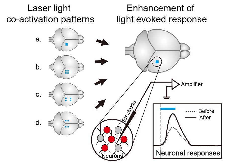
Figure legend: Left, different co-activation patterns of laser-light. Blue dots, co-activation stimulations of light-spot on the mouse brain with same total areas in different patterns. Right, enhancement of light evoked neuronal responses after co-activation. Blue dots, test light spot on the mice brain. The lower circle diagram depicts the recorded neurons and recording electrode. Neurons expressing light activated channel (ChR2) were marked red. The lower right diagram depicts the test-evoked neuronal responses before (dash line) and after (solid line) co-activation. Grey dash line, the onset of the test stimulation, blue bar, the duration of the stimulation (16.7 ms). (Image by CEBSIT)
AUTHOR CONTACT:
POO Muming
Institute of Neuroscience, Chinese Academy of Sciences, Shanghai, China
E-mail:mpoo@ion.ac.cn